How Does Titanium Mesh Support Bone Regeneration?

Time:2026-10-12 Author:Aria
0%

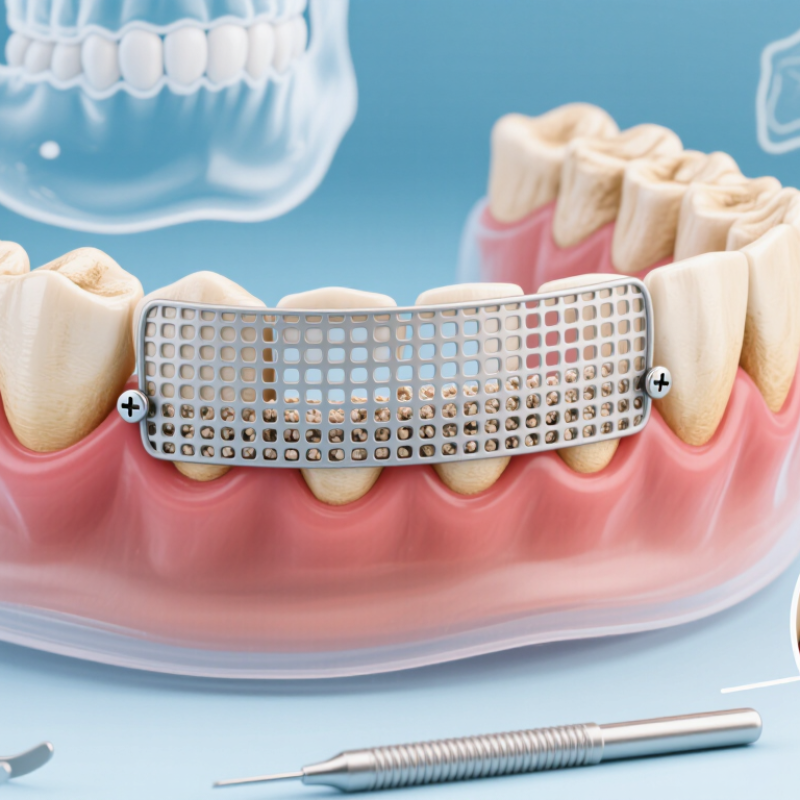
How titanium mesh supports bone regeneration is best understood through its role as a protective, space-maintaining framework. In guided bone regeneration, the mesh helps preserve a stable compartment above a bone defect. This space can hold a graft material while new bone gradually develops. Tiny openings may allow vascular access, while the rigid structure resists pressure from the gum and surrounding tissues. Think of it as a carefully shaped roof over a fragile healing site.

Clinical success depends on accurate planning and gentle handling. A dental or maxillofacial surgeon may review CBCT images, assess the defect, and contour the mesh before placement. Fixation screws help prevent movement during healing. That stability matters because repeated motion can disturb the blood clot and developing tissue. Soft-tissue closure also deserves attention. Even a well-designed mesh can cause problems if it becomes exposed.

The process is not automatic. Bone quality, defect size, smoking, infection control, graft selection, and healing capacity can influence results. In practice, outcomes vary. That fact is easy to overlook. Titanium mesh does not create bone by itself; it supports the biological conditions needed for regeneration. Its stiffness may provide dependable space maintenance, but sharp edges, exposure, or poor adaptation can compromise treatment. Therefore, evidence-based case selection and regular follow-up remain essential. Experienced clinicians must balance mechanical support with biological healing, while explaining possible complications clearly to patients. Better materials may improve handling, yet no mesh removes the need for sound diagnosis and careful surgical judgment.
How Does Titanium Mesh Support Bone Regeneration?

What Titanium Mesh Is and Its Role in Bone Repair

Titanium mesh is a thin, shaped barrier used in guided bone regeneration. It is usually made from commercially pure titanium or titanium alloy. During surgery, clinicians place it over a bone defect and secure it with small fixation screws. The mesh holds the graft in a stable contour, like a temporary wall around loose soil. This space allows blood vessels and bone-forming cells to enter gradually. It also limits soft-tissue pressure, which can otherwise flatten the graft.

Its role is mechanical, not biological. Titanium does not create bone by itself. Instead, it protects the graft while the patient’s tissues rebuild the missing volume. Systematic reviews in Clinical Oral Implants Research and the International Journal of Oral and Maxillofacial Implants commonly report implant survival above 95% after mesh-supported augmentation. However, soft-tissue exposure remains a concern, often appearing in roughly 15% to 30% of treated sites. The range varies with defect size, surgical technique, and follow-up time.

Small details matter. A poorly adapted edge may irritate the gum. Excessive tension can reduce wound stability. Careful CBCT planning, secure fixation, primary closure, and regular hygiene checks are essential. The evidence is encouraging, but it is not perfectly uniform. Some studies include few patients or short observation periods. I would not describe titanium mesh as an automatic solution. In practice, healing depends on blood supply, smoking status, infection control, and patient cooperation. Sometimes, less is safer.

How Titanium Mesh Maintains Space for New Bone Growth

How Does Titanium Mesh Support Bone Regeneration?

Titanium mesh maintains space where new bone needs to develop. After bone loss, nearby soft tissue can collapse into the defect. This leaves little room for gradual mineralization. The mesh acts like a shaped barrier and helps preserve the intended contour. It can be trimmed and adapted to the defect during surgery. Small openings allow fluid exchange while limiting unwanted soft-tissue pressure. The protected space supports a stable blood clot and may hold particulate graft material in position.

Space alone does not create bone. The defect still needs adequate blood supply, healthy surrounding tissue, and careful stabilization. Clinicians usually secure the mesh with fixation screws to reduce movement. Even slight mobility can disturb early healing. The material also resists compression from the cheek, tongue, or overlying tissue. That mechanical protection can be especially useful in wide or irregular defects.

It is not a perfect solution. Exposed mesh may irritate tissue and increase infection risk. Thin soft tissue can also make coverage difficult. For this reason, patient selection, flap handling, hygiene, and follow-up matter greatly. Healing is not automatic. A surgeon must assess the defect, the bite, tissue quality, and the patient’s medical history before choosing this approach. In practice, the best result comes from maintaining space without compromising blood supply.

How Titanium Mesh Supports Bone Regeneration

Titanium mesh helps maintain the three-dimensional space required for new bone growth. Its relatively high stiffness helps resist soft-tissue pressure and structural collapse, while its openings allow blood vessels and bone-forming cells to enter the protected defect.

Representative elastic-modulus values are shown in gigapascals (GPa). Titanium is approximately 110 GPa; cortical bone commonly ranges from about 7–30 GPa, and trabecular bone from about 0.1–2 GPa. Bone values are represented by approximate midpoints of these commonly reported ranges. Actual mesh performance also depends on thickness, pore design, fixation, and defect geometry.

How Bone Grafts and Blood Supply Support Regeneration

How Does Titanium Mesh Support Bone Regeneration?

Bone grafts and blood supply determine whether titanium mesh can support predictable regeneration. The mesh maintains a stable space above a deficient ridge. It prevents soft tissue from collapsing into the grafted area. The graft then acts as a scaffold, while new bone gradually replaces or surrounds its particles.

The blood supply is equally important. Periosteal vessels, marrow spaces, and the surrounding soft tissue deliver oxygen and repair cells. A 2023 systematic review of guided bone regeneration reported vertical bone gains commonly ranging from about 4 to 6 millimeters, although outcomes varied between studies. Exposure remains a concern, with published rates often falling between 10% and 20%. These figures are not guarantees.

Cochrane evidence on alveolar ridge preservation suggests grafting may reduce horizontal bone loss by roughly 1 to 2 millimeters during early healing. That difference can matter when implant space is limited. Yet a graft cannot compensate for poor vascular access, unstable fixation, or excessive tension in the flap. Small gaps may fill slowly. Large, dense graft masses may heal unevenly. The biology is less tidy than many diagrams suggest. Experienced clinicians therefore assess defect size, tissue thickness, systemic healing factors, and blood supply before selecting mesh-based regeneration. Titanium provides structure. Living tissue provides the regeneration.

How Does Titanium Mesh Support Bone Regeneration? — How Bone Grafts and Blood Supply Support Regeneration
Regeneration Dimension Role of Titanium Mesh Contribution of Bone Graft Importance of Blood Supply Typical Biological or Clinical Consideration
Defect-space maintenance Creates a stable three-dimensional space and helps prevent collapse of the soft tissues into the defect. Occupies the protected space and provides a scaffold or matrix for new bone formation. Allows cells, oxygen, nutrients, and signaling molecules to reach the protected region. Space maintenance is particularly important when the defect is too large for spontaneous bone fill.
Mechanical protection Provides structural support against pressure from the overlying soft tissue while the graft and early tissue are developing. Generally has limited early load-bearing capacity unless supported by native bone and fixation. Reduces the risk that poorly vascularized tissue will be damaged by excessive movement or compression. Micromotion and graft displacement can interfere with the formation of a stable provisional matrix.
Barrier function Acts as a physical barrier that can limit rapid invasion by soft tissue cells into the bone defect. Provides a protected environment in which osteogenic cells can attach, migrate, and produce new matrix. Blood vessels must still enter or connect with the grafted region for sustained cellular activity. The mesh supports guided bone regeneration but does not itself create living bone.
Osteoconduction Its framework can help contain particulate graft material and preserve the intended contour. Many graft materials provide a surface or porous structure along which new bone can grow. Vascular ingrowth is needed to support osteoblast activity and mineralized tissue formation. Osteoconduction means providing a pathway for growth; it does not necessarily mean generating new bone cells.
Osteogenesis Does not normally supply bone-forming cells or produce bone biologically. Fresh autologous bone contains living cells and may contribute directly to bone formation; other graft types primarily provide a scaffold or matrix. Survival and function of transplanted or recruited bone-forming cells depend on adequate perfusion. The biological activity of a graft depends on its source, processing, handling, particle characteristics, and local environment.
Osteoinduction Provides a protected setting but is not generally considered an osteoinductive material. Some graft matrices may contain signaling proteins that help recruit or stimulate precursor cells, although activity varies by material and processing. Vessels deliver oxygen and support the cellular responses required for signaling and matrix production. Osteoinduction and osteoconduction are different biological functions and should not be treated as equivalent.
Graft stability Can help retain particulate graft material and preserve the planned augmentation volume when properly shaped and secured. Particles or blocks must remain sufficiently stable for early tissue organization and vascular penetration. Stable tissue and vessel ingrowth are favored when the graft is not excessively compressed or mobile. Fixation method, defect geometry, soft-tissue closure, and postoperative loading all influence stability.
Blood-clot formation Helps protect the defect and maintain a contained space in which the initial clot can organize. Graft particles may become incorporated into the clot and later serve as a framework for tissue development. The clot provides an early provisional matrix; disruption can impair the transition toward granulation tissue and bone formation. Early wound stability is important because the initial clot is the foundation of subsequent healing events.
Vascular ingrowth Mesh openings allow access to the protected region, but the mesh must not excessively obstruct soft-tissue perfusion. Porosity, particle size, packing density, and graft geometry influence how easily vessels can enter the grafted area. New vessels provide oxygen, nutrients, immune-cell trafficking, and pathways for reparative cells. Large or poorly vascularized graft volumes may heal more slowly because diffusion alone cannot adequately support deep tissue.
Early inflammatory phase Provides containment while the surgical site undergoes the normal initial inflammatory response. The graft interacts with the clot and local inflammatory environment before connective tissue and bone formation progress. Inflammatory cells and soluble mediators reach the site through the circulation and help coordinate repair. Inflammation is necessary for healing, but persistent infection or uncontrolled inflammation can compromise regeneration.
Granulation tissue and matrix formation Maintains the shape and volume needed for granulation tissue to mature within the defect. Offers a surface for fibrovascular tissue organization and later deposition of osteoid. Granulation tissue is highly vascular and serves as an important bridge between the initial clot and mineralized bone. This stage commonly develops during the early weeks after surgery, although timing varies with defect size and patient factors.
Woven-bone formation Protects the developing tissue from soft-tissue pressure and helps preserve the regenerative contour. Acts as a scaffold and may be gradually surrounded or replaced by newly deposited woven bone. Active bone formation requires a continuing supply of oxygen and nutrients as cellular demand increases. Woven bone is immature and structurally less organized than mature lamellar bone.
Remodeling and maturation Can maintain the regenerated volume during early healing, but it is not intended to replace normal bone remodeling. Some graft particles may be resorbed, remain incorporated, or be progressively replaced by host bone depending on material properties and site conditions. Long-term vascular support helps coordinate osteoblast-mediated formation and osteoclast-mediated remodeling. Remodeling commonly continues for months or longer; the rate differs among individuals and anatomical sites.
Soft-tissue management Requires adequate soft-tissue coverage to reduce exposure risk and protect the regenerative compartment. Graft success depends on maintaining a closed, stable environment around the grafted area. Compromised soft-tissue perfusion can reduce oxygen delivery and impair healing. Tension-free closure, infection control, and protection from mechanical irritation are important clinical principles.
Material biocompatibility Titanium is generally considered biocompatible and can provide a rigid, corrosion-resistant framework for guided regeneration. Graft materials differ in biologic activity, resorption behavior, porosity, and interaction with host tissue. Biocompatibility helps preserve a local environment in which vascular and bone healing can proceed. Biocompatibility does not eliminate risks such as exposure, infection, inadequate fixation, or insufficient vascularization.
Overall regenerative outcome Functions mainly as a space-maintaining and protective device. Supplies a scaffold, matrix, and in some cases living cells or biologically active signals. Connects the grafted space with the body’s repair systems and supports tissue survival. Successful regeneration depends on the interaction of space, stability, graft biology, vascularity, soft-tissue coverage, and patient health.

Note: Healing time and regenerative performance vary according to defect size, anatomical location, graft characteristics, vascularity, soft-tissue condition, fixation, systemic health, and postoperative care.

How Titanium Mesh Is Positioned and Secured During Surgery

Titanium mesh supports bone regeneration by maintaining a stable space for new tissue. Its value depends heavily on accurate surgical positioning. Before surgery, clinicians review three-dimensional imaging to assess the defect, nearby nerves, tooth roots, and available bone. This planning helps determine the mesh size, shape, and fixation points.

During the procedure, the surgeon exposes the defect and places the graft material inside the planned space. The mesh is then trimmed and gently contoured to follow the patient’s anatomy. It should remain separated from delicate structures and avoid sharp edges. A stable fit matters. Even slight movement may disturb the developing graft.

Small fixation screws or pins secure the mesh to the surrounding bone. The surgeon checks that the mesh does not compress the graft or interfere with soft-tissue closure. Adequate coverage is essential because exposed mesh can increase complications. The closure should be tension-free, although achieving it is not always easy. The step that seems simplest—contouring the mesh—can create problems when rushed.

Clinical judgment remains important throughout the operation. Bone quality, defect shape, tissue thickness, and infection risk vary between patients. Follow-up imaging and examinations help confirm that the graft remains stable during healing. No single technique suits every case.

What Factors Affect Healing and Long-Term Results

How Does Titanium Mesh Support Bone Regeneration?

Titanium mesh maintains space above a deficient ridge, allowing bone-forming cells to populate the defect. Its rigidity helps resist collapse during chewing and early healing. However, long-term results depend on more than the mesh itself.

A 2023 systematic review reported average vertical bone gains of approximately 4 to 5 millimeters in selected cases. The same evidence shows membrane exposure rates commonly ranging from 20% to 30%. Exposure is not always catastrophic, but bacterial contamination can reduce the final volume. Stability matters. Even small movements may create fibrous tissue instead of mineralized bone.

Smoking, uncontrolled diabetes, poor plaque control, and thin soft tissue can delay healing. Larger defects also require longer maturation periods. Clinical teams often use CBCT imaging to assess defect geometry and monitor graft stability. The International Team for Implantology emphasizes patient selection, soft-tissue management, and strict maintenance during guided bone regeneration.

Material design also influences outcomes. A mesh with excessive rigidity may irritate tissue, while insufficient support can collapse under pressure.

Surgical experience remains important, especially when closing delicate flaps without tension.

The uncomfortable truth is that a technically correct procedure can still heal unpredictably. Some published studies include small samples and short follow-up periods. Long-term evidence remains less consistent than early bone-gain data. Careful recall visits, hygiene support, and individualized risk assessment may protect the regenerated site for years.

FAQS

What is titanium mesh used for in bone repair?

Titanium mesh is a thin, shaped barrier used during guided bone regeneration. It covers a bone defect and holds the graft in place. Think of it as a small wall around loose soil. Its role is mechanical.

How does titanium mesh help new bone grow?

The mesh preserves space above the bone defect. It limits pressure from the cheek, tongue, and soft tissue. Small openings allow fluid exchange. Space alone fails. New bone still needs blood supply and healthy surrounding tissue.

Does titanium mesh create bone by itself?

No. Titanium does not create bone biologically. It protects the graft while the body rebuilds missing bone. The graft provides a scaffold for developing tissue. Living tissue performs the actual regeneration.

Why are bone grafts important with titanium mesh?

The graft fills the defect and supports gradual bone formation. New bone can grow around or replace graft particles. A dense graft may heal unevenly. The process is less tidy than diagrams suggest.

How important is blood supply during mesh-supported regeneration?

Blood supply delivers oxygen and repair cells to the grafted area. Nearby vessels, marrow spaces, and soft tissue all contribute. Poor circulation can delay or weaken healing. The mesh cannot compensate for weak biology.

How is titanium mesh secured during surgery?

Clinicians usually trim and shape the mesh during surgery. Small fixation screws hold it against the bone. Stable fixation reduces movement during early healing. Even slight movement may disturb the blood clot and graft.

What problems can occur with titanium mesh?

Soft-tissue exposure is a common concern. Published rates often range from about 10% to 30%. A sharp edge may irritate the gum. Exposure can also increase infection risk. The exact risk varies.

What factors influence the final healing result?

Defect size, tissue thickness, flap tension, and blood supply matter. Smoking, infection control, hygiene, and medical history also influence recovery. Regular follow-up helps identify irritation or exposure early. Patient cooperation matters more than many people expect.

Is titanium mesh suitable for every bone defect?

No. A clinician must assess the defect, bite, tissue quality, and healing factors. Wide or irregular defects may benefit from its space-maintaining ability. Small defects may need a simpler approach. Sometimes, less is safer.

Conclusion

How titanium mesh supports bone regeneration begins with its role as a stable, biocompatible barrier that protects a bone graft and creates a controlled space for new tissue to form. By preventing surrounding soft tissue from collapsing into the defect, the mesh helps maintain the volume and shape needed for gradual bone growth. The graft provides a framework and mineral source, while a healthy blood supply delivers oxygen, nutrients, and regenerative cells essential for healing.

During surgery, the mesh is carefully shaped to match the defect and secured with fixation devices so it remains stable during the early healing period. Its long-term results depend on several factors, including the size and location of the bone defect, graft quality, blood circulation, infection control, soft-tissue coverage, oral hygiene, and the patient’s overall health. With appropriate planning, secure placement, and sufficient healing time, titanium mesh can support predictable bone reconstruction and create a stronger foundation for future restorative treatment.

Aria

Aria

Aria is a dedicated marketing professional with a deep passion for innovative strategies and a keen understanding of our company's product offerings. With a wealth of experience in the industry, Aria excels at crafting engaging content that highlights the unique features and benefits of our......